Dental X-rays in dental care
Radiographic examinations are essential in dentistry, as they make it possible to detect changes that are not visible to the naked eye. X-rays help diagnose cavities, gum disease, bone structure problems, and other oral conditions. The examination is quick, safe, and provides valuable information for accurate diagnosis and treatment planning.
Patients are referred for the examination by their treating physician or dentist based on a specific indication, and a referral is issued. X-rays are not performed without a referral or a clear clinical indication.
Health concerns for which diagnostic testing may be necessary
- A doctor may refer a patient for an X-ray examination for the following reasons.
- Diagnosis of dental caries: Especially between teeth or under fillings.
- Assessment of gum disease: Determining the extent of bone loss and periodontal pockets.
- Evaluation of root canal condition: Before and after root canal treatment.
- Monitoring the position and development of wisdom teeth: Before extraction.
- Planning dental implants: Assessing the volume and quality of jawbone.
- Detection of jaw cysts and tumors: Early diagnosis.
- Assessment of trauma: Identifying fractures of teeth and the jaw.
In what situations is the procedure used?
-
Comprehensive dental assessment before major treatment
If you need a comprehensive dental assessment before major treatments such as implant placement, root canal treatment or surgical procedures. -
Facial or jaw trauma
If you have sustained an injury to the facial or jaw area, for example due to a fall, impact or accident. -
Gum swelling, redness or pus
If your gums bleed, are swollen, red or show other signs that may indicate gum disease. -
Gum swelling, redness or pus
If your gums bleed, are swollen, red or show other signs that may indicate gum disease. -
Recurring inflammation
Repeated inflammation around the teeth that does not respond to treatment. -
Suspected tooth decay
If tooth decay is suspected, especially in areas that are difficult to see, such as between the teeth. -
Tooth pain, gum inflammation, or tooth sensitivity
If you experience tooth pain or tooth sensitivity that cannot be assessed based solely on external examination. -
Tooth sensitivity
If your teeth are sensitive to cold, hot or sweet foods and drinks. -
Wisdom tooth pain or inflammation
If your wisdom teeth cause pain, inflammation or do not erupt properly.
How to prepare for your appointment
- Bring an ID card or other form of identification and ensure that you have a valid referral for the X-ray examination. A Meliva doctor may also refer you directly from the treatment room, in which case a prior referral is not required.
- Remove all metal objects from the head and neck area and from the mouth (jewellery, piercings, clips, glasses, dentures). Also remove chewing gum, sweets, and food from the mouth.
What to expect during your appointment
Consultation
Your needs are discussed and the procedure is explained.
Preparation
All metal objects are removed from the head, neck, and mouth area (jewellery, piercings, clips, glasses, dentures). Chewing gum, sweets, and food are also removed from the mouth.
Protective measurese
The examined area is imaged as accurately as possible, and the rest of the body is protected with a lead apron or collar. If necessary, the apron is not used if its edge may interfere with the X-ray beam and reduce image quality or increase radiation exposure. The patient is always informed about this beforehand.
X-ray imaging
The patient stands or sits still. A bite block is placed between the teeth, and the patient is asked to keep the lips closed and the tongue against the palate. The device moves around the head. During skull lateral imaging, head supports and positioning aids are used to keep the head stable. Images are displayed immediately after the procedure.
Data processing
The images are processed and stored. X-rays taken in Meliva clinics are transferred to a national imaging database, accessible to physicians and dentists.
Result analysis
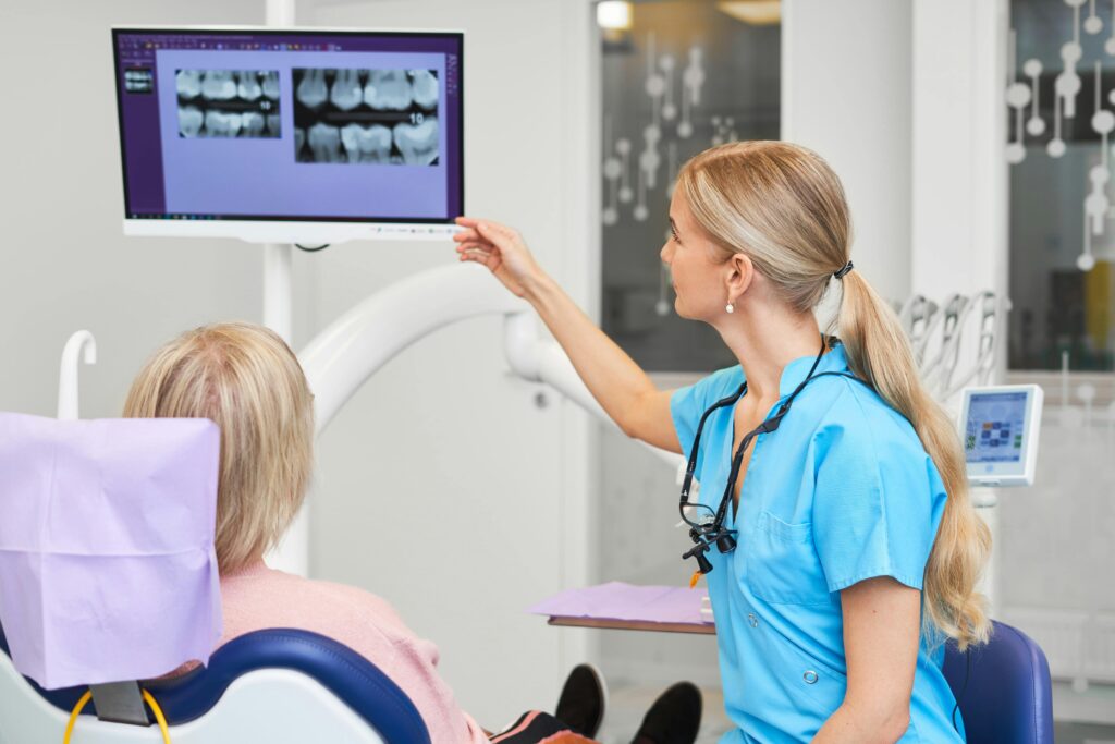
The dentist reviews the X-ray images and explains the findings.
Treatment plan
The diagnosis is explained and suitable treatment options are offered.
Payment & Appointment Info
Prices
Tallinn
| Service | Price |
|---|---|
| Visit fee in dental care Added to the price of the procedure or appointment. | 20 € |
| Specialist consultation Assessment of dental and oral health, radiographs | 70 € A visit fee is added to the price. |
| Consultation with a specialist physician | 92 € A visit fee is added to the price. |
| Panoramic or cephalometric radiograph | 32 € A visit fee is added to the price. |
| Intraoral radiograph | 19 € A visit fee is added to the price. |
| Treatment plan preparation and discussion | 68 € A visit fee is added to the price. |
| 3D X-ray scan Referral is required. | 98 € A visit fee is added to the price. |
| Head X-ray Referral is required. | 43 € A visit fee is added to the price. |
| Head X-ray – 2 images Referral is required. | 74 € A visit fee is added to the price. |
| Intraoral radiograph Referral is required. | 31 € A visit fee is added to the price. |
Tartu, Kuressaare
| Service | Price |
|---|---|
| Visit fee in dental care Added to the price of the procedure or appointment. | 20 € |
| Specialist consultation Assessment of dental and oral health, radiographs | 70 € A visit fee is added to the price. |
| Panoramic or cephalometric radiograph | 30 € A visit fee is added to the price. |
| Intraoral radiograph | 19 € A visit fee is added to the price. |
| Treatment plan preparation and discussion | 68 € A visit fee is added to the price. |
| 3D X-ray scan Referral is required. | 98 € A visit fee is added to the price. |
| Head X-ray Referral is required. | 43 € A visit fee is added to the price. |
| Head X-ray – 2 images Referral is required. | 74 € A visit fee is added to the price. |
Pärnu, Kärdla
| Service | Price |
|---|---|
| Visit fee in dental care Added to the price of the procedure or appointment. | 20 € |
| Specialist consultation Assessment of dental and oral health, radiographs | 65 € A visit fee is added to the price. |
| Panoramic or cephalometric radiograph | 29 € A visit fee is added to the price. |
| Intraoral radiograph | 17 € A visit fee is added to the price. |
| Treatment plan preparation and discussion | 65 € A visit fee is added to the price. |
| 3D X-ray scan Referral is required. | 89 € A visit fee is added to the price. |
| Head X-ray Referral is required. | 40 € A visit fee is added to the price. |
| Head X-ray – 2 images Referral is required. | 68 € A visit fee is added to the price. |
| Intraoral radiograph Referral is required. | 28 € A visit fee is added to the price. |
Payment methods
With a doctor’s referral, Meliva Health Insurance covers this service under Dental Care coverage. Check your personal coverage limits in the ERGO app.
For insured adults, the dental care benefit provided by the Tervisekassa (Health Insurance Fund) is deducted from the treatment cost at the clinic.
The Tervisekassa (Health Insurance Fund) covers the dental care for insured patients under 19 years of age.
A visit fee may be added to the cost of treatment. Familiarize yourself with Meliva’s policy on charging visit fees.
Referral
A doctor’s referral is required to book this service.
On the life long mission

Frequently Asked Questions
What is a radiographic examination in dentistry?
A radiographic examination is a diagnostic method that uses X-rays to create images of the structures of teeth, jaws, and surrounding tissues.
Why are radiographic examinations important?
X-ray examinations help detect problems that are not visible to the naked eye (e.g. cavities between teeth, root infections, bone loss, and the position of wisdom teeth).
Are X-ray examinations safe?
Yes, modern digital X-ray devices use minimal radiation doses and are safe. Modern digital radiographic systems allow images to be taken with different field sizes and parameters in order to optimize the patient’s radiation exposure. In children, special low-dose programs are used.
In Meliva clinics, we follow the ALARA (As Low As Reasonably Achievable) principle. An X-ray is performed only when there is a specific medical indication, not routinely. The benefit of the examination and the potential radiation risk are always carefully weighed. The dentist uses the lowest possible radiation dose that still provides a diagnostically sufficient and clear image.
How often should X-rays be taken?
Radiographic examinations are performed by a doctor only when there is a clear indication; X-rays are not used routinely.
What is the difference between 2D and 3D X-rays?
A 2D X-ray provides a two-dimensional image. A 3D X-ray (CBCT) provides a three-dimensional image, which is more detailed and precise. 2D images are used for general assessment, while 3D imaging is used for detailed visualization of a problem and making a definitive treatment decision.
Can X-rays be taken during pregnancy?
Radiographic procedures are generally contraindicated during pregnancy. Dental X-rays are focused on the head region, and the fetus is not exposed to the radiation field. In exceptional cases, X-rays may be performed during pregnancy if necessary. It is essential to inform the doctor and/or the technician if you are pregnant.
Are X-ray examinations painful?
No, the procedure is painless and quick.
What is 3D X-ray used for?
3D X-rays are used when a more detailed and comprehensive image of the situation is needed. A 3D scan visualizes teeth and jaws in three dimensions and allows layer-by-layer assessment. It can show, for example: inflammatory lesions, additional root canals, root fractures, or the position of the nerve canal before wisdom tooth extraction. Full jaw or whole skull 3D imaging is used for precise treatment planning in implantology, orthodontics, orthognathic surgery, etc.
Do I need to do anything before an X-ray?
Remove all metal objects from the head, neck, and mouth area (jewellery, piercings, clips, glasses, dentures). Also remove food and chewing gum from the mouth.
Is radiology expensive?
The cost of treatment depends on the type and extent of the examination. A more precise price estimate can be provided during a dental consultation. X-ray prices are also listed on the Meliva website.
What does radiation exposure mean?
Modern digital X-ray systems allow images to be taken with different sizes and parameters in order to keep the patient’s radiation exposure as low as possible. In Meliva clinics, special ultra-low-dose programs are used for children.
Meliva clinics follow the ALARA (As Low As Reasonably Achievable) principle. This means that X-rays are performed only when there is a medical indication, not routinely. Before the examination, the benefit and potential radiation risk are always assessed, and the lowest possible dose is used to obtain a diagnostically sufficient image.
The radiation is directed precisely at the examined area, and the rest of the body is protected with a lead apron or collar if necessary. In some cases, the apron may be omitted if its edge could interfere with the imaging field and affect image quality or increase radiation dose. In such cases, the technician explains this to the patient before the procedure.
What types of imaging are performed?
3D dental X-ray (CBCT)
3D dental X-ray (CBCT – Cone Beam Computed Tomography) provides a detailed three-dimensional image of the teeth and jaws. The device uses a cone-shaped beam, allowing a large number of images to be taken in a short time with a lower radiation dose.
CBCT is used when a conventional X-ray is not sufficient and a more precise overview is required. It helps visualize inflammatory lesions, root canals, root fractures, wisdom tooth position, and nerve canal location. It is also used for implant planning, orthodontic treatment, and maxillofacial surgery.
The scope and settings are determined by the dentist. The examined area may range from a single tooth to the entire skull. If needed, image-enhancing filters are used, for example to reduce motion artifacts or metal streaking, as well as ultra-low-dose (ULD) protocols for minimal radiation exposure. The examination takes about one minute, and the result is displayed immediately.
Intraoral X-ray
Intraoral X-ray is a widely used and highly precise dental examination that evaluates the condition of teeth and surrounding tissues in detail. During the procedure, a small sensor is placed inside the patient’s mouth near the area being examined.
It helps diagnose interproximal caries, decay under fillings, root infections, bone changes, gum disease, fractures, and cracks, as well as assess the need for and results of root canal treatment.
The procedure is quick and painless, although holding the sensor in the mouth may cause brief discomfort. The image is taken within seconds and displayed immediately. A protective collar for the thyroid may be used if needed.
Panoramic X-ray (PAN, PTG, OPTG)
A panoramic X-ray provides a comprehensive overview of the teeth, jaws, wisdom teeth, jaw joints, and bone structure. The examination is quick and uses a low radiation dose.
During the procedure, the patient must keep their head still, bite on a support, keep lips closed, and press the tongue against the palate. The image is taken within seconds and displayed immediately.
Lateral skull X-ray (cephalometric X-ray, CEPH)
A lateral skull X-ray is mainly used in orthodontic treatment planning and maxillofacial surgery, as well as for monitoring treatment outcomes. It allows assessment of the relationships between teeth, jaws, and facial profile, as well as jaw development.