Patient story: When knee pain becomes unbearable
Runner Irma discovered exactly why one of her knees was in such severe pain — thanks to an MRI scan.
An MRI scan is often associated with diagnosing problems in the brain or spine, but the experience of Irma Toobal, who found help at Meliva, shows that a knee problem, too, can reveal a hidden condition once you “go through the tube.” Runner Irma discovered exactly why one of her knees was in such severe pain — thanks to an MRI scan.
Before her two sons were born, Irma had run a great deal and even taken part in half marathons. Even then, she occasionally felt knee pain, but never sought help. Once her children were born, running took a back seat for close to ten years, and the knee pain faded away on its own.
At the start of last year, she set herself an ambitious goal — running a full marathon. Training was fairly intense, and the pain didn’t take long to return. “Usually, letting my legs rest brought relief, and I could get back to training. As someone who takes commitments seriously, it was mentally quite hard whenever pain forced me off my training plan,” Irma says.
At the end of August, she decided to see a physiotherapist, who gave her a set of exercises to help prevent injury. During the consultation, everything seemed fine, and she wasn’t advised against continuing to train.
Pain gives no warning — it just arrives and throws every plan into disarray
A month before the full marathon, Irma had signed up for a half marathon, and the day before that, a 10km fun run. She treated the fun run as more of a light training session, with no plans to set any records — just to enjoy the atmosphere.
Although her knee felt oddly tight that morning, the feeling was gone by evening, and Irma went out to run. “The run went fine, but the next morning I woke up with such severe knee pain that I couldn’t put any weight on it. Naturally, I didn’t make it to the half marathon that evening either,” the amateur athlete recalls. “I was really shaken, because I realised I couldn’t even hop on one leg to the pharmacy to buy painkillers. At home, I got around by pushing a chair in front of me and hopping on one leg. I spent that whole day and the next in bed, but the pain didn’t ease.”
This turn of events came as a real shock to Irma, since she hadn’t suffered any direct injury to her leg, and everything had felt fine when she went to bed the night before. Overnight, though, her knee had swollen up considerably. So she rented crutches and quickly booked an appointment with Meliva orthopaedist Mart Pais.
“The pain was still severe, and I couldn’t put weight on the leg at all — it simply wouldn’t hold me. By the time I reached the doctor, I’d just taken a fairly large dose of painkillers, which meant I was able to get through the necessary tests. On examination, nothing seemed visibly wrong with the knee, and the cause of the pain remained unclear. Since I still couldn’t manage without painkillers, the issue needed further investigation.”
How did an MRI reveal what was behind the knee pain?
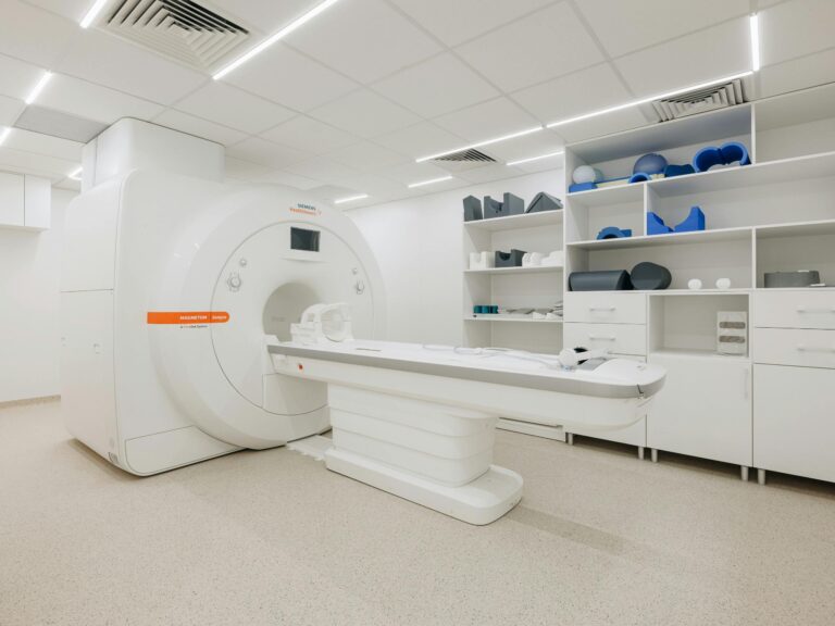
The orthopaedist referred Irma for an MRI (magnetic resonance imaging) scan at Meliva’s Rävala clinic, which allows very precise examination of different areas of the body using a magnetic field and radio waves. An MRI machine is suited to examining bones, joints, the brain and spinal cord, and, with some exceptions, the abdominal and pelvic organs. Irma recalls going into the “tube” only up to her hips. Before entering the machine, the radiology technician helped her get into as comfortable a position as possible, since she needed to lie completely still for 20 minutes.
Before the results came back, though, the marathon Irma had spent eight months training for arrived. Her doctor had advised her to stick to cycling and swimming in the meantime, to maintain fitness while avoiding putting excess strain on her knees. On the eve of the marathon, though, she decided to take part anyway, promising herself she wouldn’t be hard on herself and would drop out — even in the very first kilometres — if the pain became unbearable.
So Irma loaded up on painkillers, magnesium, and sports gel before the start, and set off. Her nerves were running high, adrenaline was through the roof, and she couldn’t manage to eat a thing. The first seven kilometres passed in a battle against nausea, but her legs, at least, were holding up well.
“I decided to keep going anyway. The nausea passed, and I felt really good all the way up to the 36th kilometre. From there, there was so little left (a full marathon is 42.195 km long — editor’s note) that I couldn’t bring myself to stop. I alternated between walking and running all the way to the finish. My soles were burning, my legs ached — but not in the knees, elsewhere. My family and friends supported and cheered me on the whole way round. I saw pure pride in my sons’ eyes as they watched me cross the finish line. I was overjoyed to have finished the marathon! Never mind that it took an hour longer than I’d planned.”
A harsh truth after the marathon
Eventually, the MRI results came back — and they were anything but reassuring. It turned out Irma’s knee had a pre-stress-fracture condition. Looking back, Irma says that if she’d seen Dr Pais earlier and gotten a diagnosis before the race, she certainly wouldn’t have run. “My story shows that no matter how good a doctor is, they can’t see fully inside us or make a precise diagnosis without the right equipment. Even though my knee seemed to be functioning fine on the surface and the painkillers were working, the doctor still decided to investigate further. I’m very grateful to Dr Pais for taking my concern seriously,” she says. “An MRI and X-ray are the ideal tools for finding out what’s actually going on inside our bodies,” she adds.
Recovery after the marathon took three months, and only then could Irma start running shorter distances again. A follow-up X-ray, taken on the orthopaedist’s recommendation, confirmed the condition was improving and that the marathon hadn’t actually caused a full fracture. “I still avoid more serious running for now, and prefer cycling, swimming, and long walks instead.” Still, Irma admits in closing, her heart still belongs to running.
Meliva orthopaedist Dr Mart Pais:
A young woman in good athletic shape came in on forearm crutches, since putting weight on her leg was painful. The clinical examination (a physical assessment using sight and touch) didn’t reveal anything conclusive, and based on the patient’s history, an X-ray wasn’t indicated either.
The MRI scan revealed, unexpectedly, a pre-stress-fracture condition in the lower femur, in the area of the knee joint, showing up as bone swelling. MRI distinguishes tissue based on fluid content, whereas changes visible on an X-ray only appear over time and often point to an already more serious situation.
Research shows that stress fractures account for roughly 20% of all sports injuries. Runners who cover more than 40 km a week face a higher risk. The most common sites for stress fractures are the shinbone, the navicular bone in the foot, the metatarsal bones, the femoral neck, and the pelvic bones.
Stress fractures can sometimes point to an underlying, previously undiagnosed condition. That was the case for Irma too — blood tests helped identify the root cause of the problem and start the right treatment.
Often, a precise diagnosis can be reached through the patient’s account (their history), a clinical examination, and, where needed, an X-ray. That said, improved access to MRI has made faster diagnosis and more precise answers possible, helping serve patients more effectively.